Museum celebrates RPH role in HIV research race

A very special exhibit has opened at the Royal Perth Hospital (RPH) Museum – one that encapsulates the primary role played by the hospital in the discovery of HIV in human tissue.
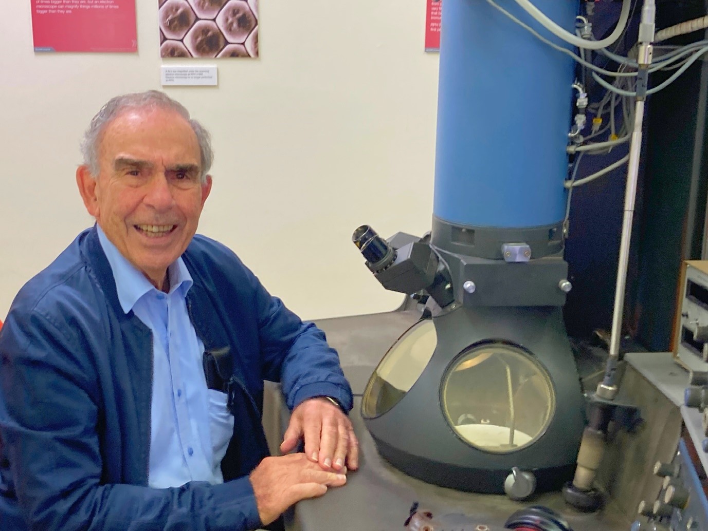
As part of the Boorloo Heritage Festival which ran over the weekend of April 15th, the RPH Museum showcased the transmission electron microscope (TEM), which remains in its original position since it was first commissioned in 1980 at the old hospital building.
It was using this microscope that Dr John Armstrong and Mr Robert Home of the RPH Department of Anatomical Pathology along with their dedicated team won the global race to demonstrate the presence of virus particles in patients with HIV infection.
Dr Armstrong captured the first image of the virus particles in human tissue, something which ultimately shaped the way AIDS was treated and diagnosed across the world.
Dr Armstrong made the landmark discovery in 1984 and published his findings in the world-renowned journal The Lancet, which attracted acclaim from thousands of healthcare workers and scientists across the globe – and he did all of this while working at RPH.
Earlier this week, Professor John Papadimitriou – who worked with Dr Armstrong and on the TEM in the 1980s – returned to RPH for a look at the ground-breaking microscope, before the exhibit becomes crowded by curious viewers over the weekend.
Reminiscing about the days he worked on the TEM, Prof Papadimitriou recalled the moment Dr Armstrong discovered the virus particles in human tissue, calling him a “celebrity” in the science world.
“I was there when he spotted HIV in human tissue in 1984, and he was extremely excited. All of a sudden, the whole theory started to make sense behind HIV,” Prof Papadimitriou said.
“In those days, there were several theories behind HIV and the causation of AIDS. But seeing the virus particles and then incorporating the other data that had been collected from around the world, like France and the US, everything came together.
“This was the first microscope in the world to find HIV in human tissue. That was achieved by Dr Armstrong and fellow pathologist Dr Robert Home, when they saw the virus particles in human samples, and soon after, were able to demonstrate that this was in fact HIV,” he added.
Prof Papadimitriou said this discovery at RPH “enabled scientists and medical practitioners to develop a therapy for the disease, which was plaguing parts of the world.”
“Dr Armstrong became a bit of a celebrity after the discovery. Throughout the world, people were reaching out to him and our office to get more data or just simply discuss his findings – it was amazing,” he remembered fondly.
“This was ground-breaking because by knowing what tissues the virus affected and by knowing what the virus does and how it does it, medical scientists were able to develop appropriate therapies to treat people with AIDs.
“Dr Armstsong’s findings were at the forefront of how AIDs was treated. Royal Perth was essentially leading the globe in this development,” Prof Papadimitriou said.
“Using this TEM, we were also the first people in the world to photograph helicobacter pylori, which is a type of bacteria that lives in the lining of the stomach,” he added.
Come down and speak to some of RPH’s knowledgeable museum staff between 9am and 1pm every Wednesday and Thursday, to get the full history of the TEM and learn about its role in the trailblazing research that occurred back in the 1980’s.

